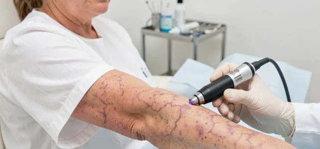
A bluish network on the leg, a marbled appearance that appears after a shower or when it’s cold: this type of skin marbling is common and, most of the time, not serious. Marbled skin is concerning because it visually resembles certain pigmented lesions. Knowing how to distinguish a simple reaction to cold or dehydration from a skin sign that requires medical advice helps avoid both unnecessary anxiety and diagnostic delays.
Physiological livedo and cutis marmorata: the vascular mechanism behind marbled skin
Have you ever noticed a bluish network on your thighs after getting out of a warm bath? This phenomenon has a name: cutis marmorata. It is a reflex contraction of small blood vessels under the skin in response to cold.
The pattern is symmetrical, regular, and disappears within minutes as the skin warms up. It causes no pain, itching, or peeling. This physiological livedo mainly affects the legs and arms, and it is more visible on lighter skin.
Dehydration accentuates this marbled effect. When the body lacks water, microcirculation slows down, and blood stagnates more in the superficial venules. The skin fold test, described notably by Michel Cymes on RTL, allows for a quick check of hydration levels: pinch the skin on the back of the hand and observe if the fold retracts in less than two seconds. A persistent fold indicates dehydration, which can explain a temporary marbled appearance on its own.
To better understand the distinction between dehydration marbled skin and cancerous spots, three simple criteria should be observed: the symmetry of the network, its disappearance upon warming, and the absence of raised lesions.

Persistent and asymmetrical marbling: when the skin signals something other than a chill
A livedo that does not disappear upon warming warrants medical advice. This is the clearest line of demarcation between a benign phenomenon and a pathological sign.
Several elements should raise concern:
- The marbled network is asymmetrical, irregular, and affects only one limb or a localized area of the body.
- The marbling persists for several hours, even in a warm environment, and is accompanied by pain, a burning sensation, or ulcerations.
- Other symptoms appear in parallel: unusual fatigue, fever, joint pain, or a history of venous thrombosis.
This type of persistent livedo, described in the medical literature as pathological livedo reticularis, can reveal a systemic vascular disorder. The antiphospholipid syndrome (APS) is a documented example: this coagulation disorder causes skin marbling that can sometimes be the very first visible sign of the disease, even before thrombotic episodes.
This vascular cause is rarely mentioned in public content about marbled skin, even though it represents a differential diagnosis to be aware of so as not to attribute everything to skin cancer.
Cancerous spots on the skin: visual markers that separate them from marbling
Potentially cancerous skin lesions do not resemble a venous network. They appear as isolated spots, nodules, or altered moles. The confusion arises because some dark pigmented lesions can, from a distance, resemble a marbled area.
The ABCDE rule applied to suspicious lesions
This self-monitoring method, used by dermatologists for melanoma screening, is based on five criteria:
- A for Asymmetry: the lesion is not round or oval; one half differs from the other.
- B for Irregular Borders: the edges are jagged, blurry, or poorly defined.
- C for Heterogeneous Color: multiple shades coexist within the same lesion (brown, black, red, white).
- D for Diameter: the lesion exceeds the size of a pencil eraser.
- E for Evolution: the size, shape, color, or thickness changes over a few weeks or months.
Marbled skin, even if visually impressive, does not meet any of these criteria. The venous network has no clear border, no localized heterogeneous color, and does not form a palpable relief.

Basal cell carcinoma and often ignored lesions
Melanoma is not the only skin cancer. Basal cell carcinoma, the most common form of skin cancer, often begins as a small translucent or pearly bump, sometimes crossed by small visible vessels. It can also appear as a sore that does not heal for several weeks.
A sore that does not heal within three weeks justifies a dermatological consultation, even if it does not resemble the classic image of a suspicious mole. This criterion is more reliable than color alone for spotting an early carcinoma.
Marbled skin in infants and adults: two different contexts
In infants, marbled skin (cutis marmorata) is an almost universal phenomenon. The vascular system is still immature, and marbling appears at the slightest change in temperature. It disappears spontaneously in the first months of life, without treatment.
In adults, the situation is different. A livedo that gradually develops after years without visible marbling, or that worsens, warrants investigation. The doctor will first look for a vascular or autoimmune cause before considering a link to malignant pathology.
Marbled skin is not, in itself, a sign of cancer. It reflects a circulatory phenomenon. It is the localized, raised, evolving, or ulcerated lesions that point towards an oncological diagnosis.
The most useful reflex remains regular monitoring of one’s skin, ideally every three months in front of a mirror. Any new lesion that persists, changes, or bleeds should be shown to a dermatologist, regardless of whether it appears marbled or not. The timeliness of diagnosis remains the most significant factor influencing the prognosis of skin cancers.